
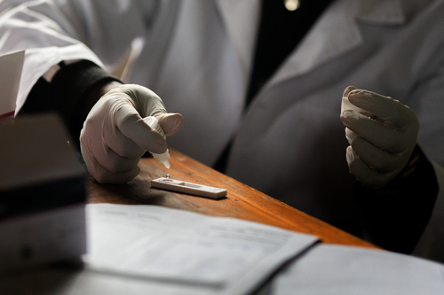
Recognising helminth infections: why diagnosis matters
Practical clinical considerations for healthcare professionals, with a focus on soil-transmitted helminths and schistosomiasis in sub-Saharan Africa.
Helminth infections can enter the consultation through many different complaints: abdominal discomfort, anaemia, blood in urine, recurrent diarrhoea or genital symptoms. Some patients have few symptoms; others present with complications. Recognition begins by connecting the clinical picture with exposure and then choosing an investigation capable of answering the relevant question.
For healthcare professionals, diagnosis matters because “worms” are not a single condition. Parasites differ in their transmission, the specimens required to detect them, their complications and their treatment. A generic stool examination or a previous course of deworming cannot answer every clinical question.
This article focuses on common intestinal soil-transmitted helminths, Strongyloides stercoralis and schistosomiasis. It brings together practical considerations for history-taking, investigation and follow-up. Test availability and care pathways vary, so the approach should be adapted to national guidance and local laboratory capacity.
Start with exposure, including exposure long before presentation
A useful exposure history goes beyond the country where the patient currently lives. Ask about previous residence, the communities visited and the activities undertaken there. Relevant details include freshwater contact through bathing, swimming, fishing or work; contact with potentially faecally contaminated soil; sanitation conditions; and previous antiparasitic treatment.
Record the approximate timing of exposure and symptom onset. Diagnostic yield can depend on the stage of infection: early symptoms or eosinophilia may occur before mature intestinal worms produce eggs detectable in stool. CDC’s clinical guidance also emphasises that particular exposures and incubation periods help narrow the differential diagnosis. [1]
For strongyloidiasis, remote exposure remains relevant because autoinfection can sustain infection for years. A patient who left an endemic area long ago may still require assessment, particularly before immunosuppression. [2]
Make the history specific enough to guide the laboratory request. “Previous freshwater contact in an endemic area” is more useful than “travel abroad”. Likewise, document the name and date of previous treatment where possible, rather than assuming that all deworming regimens covered the same parasites.
Recognise patterns, while keeping a broad differential
Symptoms suggest where to investigate; they rarely establish the diagnosis alone. The following patterns are prompts for assessment rather than diagnostic criteria.
| Presentation in a patient with relevant exposure | Helminth infection to consider | Useful next step |
|---|---|---|
| Iron-deficiency anaemia, sometimes with abdominal symptoms | Hookworm | Investigate anaemia and request appropriate stool parasitology. [3] |
| Persistent diarrhoea, particularly with heavy infection and rectal prolapse in a child | Trichuris trichiura | Assess severity and hydration; arrange stool microscopy. [4] |
| Abdominal symptoms, or possible intestinal or biliary obstruction | Ascaris lumbricoides | Request stool investigation in stable patients; investigate obstruction urgently when suspected. [5] |
| Haematuria or urinary symptoms after freshwater exposure | Urogenital schistosomiasis | Arrange urine examination for eggs and assess other causes of haematuria. [6,7] |
| Intestinal symptoms with hepatosplenic findings after freshwater exposure | Intestinal schistosomiasis | Request stool investigation and assess possible organ complications. [6,7] |
| Recurrent gastrointestinal complaints with urticaria or a rapidly moving serpiginous rash | Strongyloidiasis | Request Strongyloides-specific assessment. [8] |
| Genital symptoms with relevant freshwater exposure | Genital schistosomiasis | Undertake appropriate genital assessment and evaluate competing diagnoses. [9] |
Avoid attributing anaemia, diarrhoea or weight loss to helminths solely because exposure is plausible. A positive parasite finding may coexist with another condition; it does not necessarily explain every symptom. The clinical task is to establish both the infection and the cause of the presenting problem.
Use eosinophilia as a clue, not a gatekeeper
An increased absolute eosinophil count can support suspicion of a helminth infection, but it is nonspecific. Allergic disease, medication reactions and other conditions can also cause eosinophilia. Use the absolute count rather than the percentage alone, and interpret it alongside the history and examination. [1]
The absence of eosinophilia should not end an otherwise justified investigation. Immune responses vary, and eosinophilia may be absent in severe strongyloidiasis. [8] Conversely, eosinophilia does not identify the parasite or quantify the severity of disease.
In practice, the blood count is part of the assessment rather than a substitute for parasite-specific testing. When eosinophilia is unexplained, revisit exposures and medications and discuss appropriate investigations. When exposure is relevant but the eosinophil count is normal, choose testing according to the clinical question and risk.
Intestinal helminths: ask what the stool test actually detects
Stool microscopy can detect eggs of Ascaris, Trichuris and hookworms. Hookworm infection can cause intestinal blood loss and iron-deficiency anaemia, especially with heavier infection. [3] Heavy trichuriasis can produce persistent diarrhoea and rectal prolapse, while ascariasis can cause intestinal obstruction or complications when worms enter the biliary tract. [4,5]
These differences matter when interpreting a presentation. An uncomplicated positive stool examination and a patient with an acute obstructive syndrome require different management pathways. A stool request should not delay investigation or stabilisation of a clinically unwell patient.
Check which method the local laboratory uses. A report labelled “stool examination” does not necessarily mean that the sample was examined using a concentration technique, a quantitative egg-counting method or a method suitable for Strongyloides. Communicate the organisms suspected and the reason for the request.
A meta-analysis found that sensitivity varies with parasite species, method and infection intensity. Additional samples or slides can improve detection; light infections are more easily missed. [10] Collection instructions, correct identification and adherence to the laboratory’s specimen requirements therefore contribute directly to diagnostic quality.
When reviewing a negative report, ask whether it came from a single specimen and whether the method was appropriate. A result should be interpreted as the outcome of a specific investigation, not as evidence that all helminth infections have been excluded.
Schistosomiasis: match the specimen to the likely species
Freshwater exposure in an endemic area is central to the assessment. Intestinal schistosomes, including Schistosoma mansoni, shed eggs in stool; S. haematobium sheds eggs in urine. [19] In some parts of sub-Saharan Africa both occur, so investigating both stool and urine may be appropriate. CDC recommends collecting three samples on different days to improve microscopy sensitivity. [7]
Haematuria supports suspicion of urogenital schistosomiasis but does not establish an individual diagnosis. A urine dipstick is not a sufficiently sensitive or specific replacement for appropriate investigation in an individual patient. [7] Persistent haematuria also warrants assessment for other urinary pathology.
Schistosomiasis may cause disease even when symptoms are limited. Intestinal and hepatosplenic disease, urinary tract damage and genital involvement require attention beyond detecting eggs. [6] Where examination suggests complications, additional clinical investigations and referral should proceed according to the organ involved and local capacity.
Early negative investigations after a recent exposure require particular care. The timing of infection affects what can be detected. Document the exposure date and arrange reassessment rather than treating an early negative test as definitive reassurance.
Understand what antibody, antigen and molecular tests can tell you
Schistosoma antibody testing can support diagnosis in previously untreated travellers or migrants with relevant exposure. For a new infection, CDC advises obtaining the serum sample at least six to eight weeks after likely exposure. Antibodies may persist after successful treatment, limiting their ability to distinguish past from active infection in repeatedly exposed or previously treated patients. [7]
Antigen tests and molecular methods offer additional approaches, but their usefulness depends on the species, assay and setting. Antigen detection can provide evidence of active infection; assay performance and interpretation are not interchangeable. A recent review describes the strengths and limitations of microscopy, serology, antigen detection and molecular diagnosis. [11]
Before ordering a less familiar test, establish what question it answers. Is the assay intended to support individual diagnosis, screen a population or monitor a programme? What does a positive result mean after treatment? What confirmatory or follow-up investigation is available?
When a result does not fit the clinical picture, discuss it with the laboratory or a clinician experienced in parasitic disease. The appropriate response may be another specimen, a complementary method or reassessment of the differential diagnosis. It should not depend on a simple hierarchy in which a newer technology is assumed to be definitive.
Strongyloidiasis needs a dedicated diagnostic strategy
Strongyloidiasis is frequently asymptomatic or produces nonspecific gastrointestinal and skin symptoms. Larva currens—a recurrent, rapidly advancing serpiginous rash, often affecting the buttocks, perineum or thighs—is an important clinical clue. Severe disease can involve the gastrointestinal tract, lungs and other organs. [8]
Routine stool examination may miss infection because larval output is low or intermittent. Strongyloides assessment may require serial specimens, specialist larval detection techniques, serology or molecular testing, depending on local availability. Serology is useful but can be less sensitive in immunocompromised patients and requires contextual interpretation. [2]
The laboratory request should explicitly name Strongyloides when it is suspected. Confirm whether the service can perform an appropriate test or needs to send the specimen elsewhere. A negative routine examination for parasite eggs is insufficient reassurance in a high-risk patient.
Evidence-based guidance supports using sensitive serology for screening and combining serological and parasitological methods in immunosuppressed patients where possible. Screening should occur before immunosuppression when feasible, because subsequent immune suppression can affect test sensitivity. These guidelines were developed for non-endemic countries; their application in endemic settings should be aligned with local protocols. [12]
Before immunosuppression, make the Strongyloides question explicit
Patients with relevant exposure who are about to receive corticosteroids or other immunosuppressive treatment need particular attention. Other important contexts include transplantation, haematological malignancy and HTLV-1 infection. CDC specifically highlights recent or remote endemic-area exposure in its screening considerations. [13]
An applicable clinical sequence is to document exposure, establish whether adequate previous treatment is recorded and arrange assessment through the agreed local pathway. If immunosuppressive treatment is urgent, seek prompt specialist advice rather than allowing routine test turnaround to determine management.
Guidelines allow consideration of pre-emptive treatment in selected high-risk situations when appropriate testing is unavailable, particularly before immunosuppression. [12] This is a clinical decision requiring evaluation of treatment risks. Confirmed or suspected Loa loa coinfection is a relative contraindication to ivermectin, and pregnancy, lactation and low body weight also require specific consideration. [13]
The aim is to anticipate risk at the treatment decision, not to wait for deterioration. Record who will review outstanding results and how they will be communicated to the team prescribing immunosuppression.
Genital schistosomiasis belongs in the differential diagnosis
Female genital schistosomiasis can present with pelvic pain, dyspareunia, vaginal discharge or contact and postcoital bleeding. These symptoms overlap with sexually transmitted infections and other gynaecological conditions. WHO stresses the need to assess competing diagnoses, including malignancy and urogenital tuberculosis. [9]
Relevant freshwater exposure should prompt consideration of genital involvement, even when urinary investigations do not confirm infection. Laboratory detection of eggs and recognition of genital disease are not equivalent: eggs may not be demonstrable alongside the genital lesions. [9]
WHO’s Female Genital Schistosomiasis Pocket Atlas provides visual support for trained healthcare professionals working in endemic, low-resource settings. It can assist recognition during appropriate examination, while abnormal or uncertain findings still need the relevant referral pathway. [14]
Discuss the possibility sensitively. Schistosomiasis is acquired through contaminated freshwater, and genital symptoms should not automatically be attributed to sexual transmission. Assessment for sexually transmitted infections and other conditions should continue where indicated; one diagnosis does not exclude another.
Recognise when routine outpatient investigation is insufficient
Some presentations require urgent assessment and management regardless of whether a parasite has been confirmed. Examples include suspected bowel obstruction, major bleeding, severe symptomatic anaemia, respiratory failure, sepsis or new neurological deficits. These are indications to assess and stabilise the patient, with parasitic infection considered alongside other possible causes.
In an exposed patient who has received corticosteroids, gastrointestinal and respiratory deterioration, particularly with enteric bacterial sepsis, should raise concern for Strongyloides hyperinfection. [8] Urgent specialist and laboratory involvement is needed; waiting for a routine outpatient stool result is inappropriate in this setting.
Schistosomiasis can also produce serious complications, including hepatosplenic and urinary disease. [6] The diagnostic plan should assess both infection and organ damage. Finding eggs may clarify aetiology, but it does not replace the investigation of a complication already suggested by examination or imaging.
When arranging referral, provide the exposure history, medications, previous treatment, test methods and available results. This information helps the receiving team choose the next investigation without unnecessarily repeating the assessment.
A negative test needs a clinical interpretation
The key question is whether the investigation was capable of detecting the suspected infection under the circumstances. Review the specimen, method, timing and number of samples. For low-intensity infections, a single negative microscopy result may leave substantial uncertainty. [10,11]
If suspicion remains, agree a specific next step: repeat sampling, complementary testing, specialist advice or assessment for another diagnosis. Repeating the same low-yield investigation without considering its limitations is unlikely to resolve the uncertainty efficiently.
Explain the result clearly to the patient. “No eggs were seen in this specimen” communicates the scope of microscopy more accurately than an assurance that no parasitic infection is possible. Give collection instructions for further samples and identify how the patient will receive the outcome.
The same principle applies to positive findings. Consider whether the parasite detected explains the symptoms, whether coinfection is plausible and whether further assessment is needed. Laboratory confirmation strengthens clinical reasoning; it should not prematurely close it.
Diagnosis should lead to treatment and a follow-up plan
Treatment needs to match the organism and patient circumstances. Evidence shows that commonly used anthelmintics differ in efficacy against Ascaris, hookworm and Trichuris. [15] A previous deworming dose does not establish that every plausible infection was treated successfully.
For schistosomiasis, praziquantel treatment and its timing require attention to species, exposure and infection stage. CDC recommends follow-up stool or urine examination one to two months after treatment when eggs were demonstrable before treatment. [16] For Strongyloides-positive patients with persistent symptoms, CDC recommends follow-up stool examinations two to four weeks after treatment. [13] Other follow-up approaches may be appropriate under specialist or national guidance.
Persistent symptoms require reassessment for treatment failure, reinfection, complications and alternative diagnoses. Symptom improvement alone is not a universal test of parasitological cure, and persistent antibodies do not necessarily establish ongoing active schistosomiasis. [7]
Provide practical advice on reducing future exposure where possible, recognising the limits imposed by local water and sanitation access. Ensure that the treatment record and follow-up plan remain available if the patient attends another service.
Individual care and preventive programmes are complementary
Preventive chemotherapy programmes use population-level criteria to reduce morbidity. WHO’s schistosomiasis guideline addresses these interventions alongside measures to reduce transmission. [17] Their purpose differs from investigating a symptomatic patient or assessing risk before immunosuppression.
Participation in a programme should be documented, but it should not replace individual clinical assessment when symptoms persist or a high-risk decision is imminent. Ask what was administered and when, then determine whether additional investigation is justified.
DIDIDI is developing affordable digital diagnostics and connected information systems for underserved rural communities, with planned evaluation of accuracy, usability and deployment. [18] Such research aims to strengthen future diagnostic capacity. In current practice, healthcare professionals can improve care by asking specific exposure questions, choosing appropriate tests and ensuring that results lead to action.
A practical approach at the consultation
Begin with the presenting syndrome and assess its severity. Document relevant freshwater, soil and residential exposure, including remote exposure where Strongyloides is possible. Select the specimen and test for the parasite suspected, and tell the laboratory what you are investigating.
Interpret the result alongside the method and timing. If uncertainty remains, make the next step explicit rather than leaving an unresolved negative report in the record. Check for complications, use parasite-specific treatment guidance and arrange follow-up appropriate to the infection and patient.
Recognition is the first step; a complete diagnostic pathway carries that recognition through to safer clinical care.
References
-
Wendt EM, Kamb ML, Montgomery SP. Post-Travel Parasitic Disease Including Evaluation of Eosinophilia. CDC Yellow Book, 2026 edition. CDC.
-
Buonfrate D, Bradbury RS, Watts MR, Bisoffi Z. Human strongyloidiasis: complexities and pathways forward. Clinical Microbiology Reviews. 2023;36(4):e00033-23. doi.org/10.1128/cmr.00033-23.
-
Centers for Disease Control and Prevention. DPDx—Intestinal Hookworm. CDC.
-
Centers for Disease Control and Prevention. DPDx—Trichuriasis. CDC.
-
Centers for Disease Control and Prevention. DPDx—Ascariasis. CDC.
-
Centers for Disease Control and Prevention. Clinical Overview of Schistosomiasis. CDC.
-
Centers for Disease Control and Prevention. Clinical Testing and Diagnosis for Schistosomiasis. CDC.
-
Centers for Disease Control and Prevention. Clinical Overview of Strongyloides. CDC.
-
World Health Organization. Schistosomiasis: Genital manifestations. Questions and answers. 2020. WHO.
-
Nikolay B, Brooker SJ, Pullan RL. Sensitivity of diagnostic tests for human soil-transmitted helminth infections: a meta-analysis in the absence of a true gold standard. International Journal for Parasitology. 2014;44(11):765–774. PubMed.
-
Ally O, Kanoi BN, Ochola L, et al. Schistosomiasis diagnosis: Challenges and opportunities for elimination. PLOS Neglected Tropical Diseases. 2024;18(7):e0012282. doi.org/10.1371/journal.pntd.0012282.
-
Requena-Méndez A, Buonfrate D, Gomez-Junyent J, Zammarchi L, Bisoffi Z, Muñoz J. Evidence-Based Guidelines for Screening and Management of Strongyloidiasis in Non-Endemic Countries. American Journal of Tropical Medicine and Hygiene. 2017;97(3):645–652. doi.org/10.4269/ajtmh.16-0923.
-
Centers for Disease Control and Prevention. Clinical Care of Strongyloides. CDC.
-
World Health Organization. Female genital schistosomiasis: A pocket atlas for clinical health-care professionals. Geneva: WHO; 2015. WHO.
-
Moser W, Schindler C, Keiser J. Efficacy of recommended drugs against soil transmitted helminths: systematic review and network meta-analysis. BMJ. 2017;358:j4307. doi.org/10.1136/bmj.j4307.
-
Centers for Disease Control and Prevention. Clinical Care of Schistosomiasis. CDC.
-
World Health Organization. WHO guideline on control and elimination of human schistosomiasis. Geneva: WHO; 2022. WHO.
-
DIDIDI consortium. Project description. DIDIDI.
-
Centers for Disease Control and Prevention. DPDx—Schistosomiasis Infection. CDC.
Sources accessed on 13 October 2026. This clinical overview focuses on selected helminth infections; testing, treatment and referral should follow applicable national guidance and local specialist advice.